A generous grant from The John & Birthe Meyer Foundation for radiation therapy equipment at DTU has paved the way for new breakthroughs with the potential to improve radiation treatment of cancer.
Radiation therapy is an effective way to treat cancer and can often cure it completely. It involves bombarding the cancerous tumour with powerful x-rays, thus destroying the hereditary material in the cells—which is usually sufficient to kill them.
"The new equipment at DTU will provide an important source of reference when we’re developing the particle radiation equipment."
Professor Cai Grau, MD, the Department of Oncology at Aarhus University Hospital
In this regard, accuracy is the key
The rays are lethal by their very nature; not only to the cancerous tumour, but also to the healthy tissue they pass through en route. For this reason, accuracy is crucial when planning and performing modern radiation therapy. It is both a matter of accurately hitting the cancerous growth, and administering precisely the dose required to kill the tumour, neither more nor less.
In order to determine the precise dose for a given treatment, it is initially necessary to ensure that the radiation equipment itself is ideally suited to the operation and properly calibrated. Moreover, it is essential to have a complete picture of the patient and the exact location of the tumour. Such pictures are normally prepared using data from CT and MRI scans, which illustrate how the radiation disperses its energy.
 |
|
With a new radiation cannon, DTU researchers can develop much more accurate methods for measuring the doses administered to patients.
It can also be used in hospitals to verify specific treatment plans and to develop new forms of treatment.
|
Scanning and radiation equipment
The latest developments in the field of scanning and radiation equipment are opening the door to increasingly complex treatments. Before the new treatments can be utilized in practice and applied to human patients, however, comprehensive measurements have to be conducted to document that they can be performed with the greatest safety. The problem is that the dosimetry (the measurement of ionizing radiation) and the methods for performing such measurements in a clinical environment have not kept pace with technological developments. That is why new and improved measurement methods are needed.
A donation of almost DKK 19 million from The John & Birthe Meyer Foundation has now allowed DTU Nutech to purchase an accelerator on a par with the very best hospital model, and a cobalt-60 reference unit with associated electronics of the finest calibre.
“This puts us in a unique position to increase our knowledge about dosimetry, and our aim is to become a national competence centre in the field. Our ultimate goal is to help hospitals offer patients the best and most modern radiation therapy,” says Claus Andersen, Senior Scientist at DTU Nutech, which, for the past ten years, has been working extensively with doctors and hospital physicians in both methods and equipment for dosimetry.

Accelerator and cobalt source
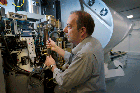
Behind a solid safety door and an intricate system of alarms—in a room where the ventilation system replaces 21,000 m3 of air every hour, making it possible to keep the temperature completely stable—stands the new Varian TrueBeam electron accelerator, an ultra-modern radiation cannon.
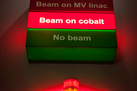
In addition to the accelerator, DTU has purchased a cobalt source. The cobalt 60 isotope always emits the same volume of energy, so it is common to calibrate dosimetry instruments—known as ion chambers—in a cobalt 60 installation. However, a great deal of calculation work must then be performed to establish how to transfer the measurement to the accelerator beam used in a given hospital.
“DTU now has both a cobalt source and an accelerator, so conditions are perfect for being able to calibrate directly in the accelerator beam, which will provide much more accurate knowledge about the radiation dose in a specific treatment,” adds Claus Andersen.
Ambitious goals
The basis for all measurement technology is to have internationally recognized primary standards. Work in this field is coordinated by an international standards laboratory in Paris. The aim is for DTU to contribute to this work and to develop a separate primary standard for radiation dosimetry.
“Our schedule is to have both the cobalt source and the accelerator accredited within two years, as we aim to draw traceability from an overseas standard laboratory. In no more than five years, we hope to receive accreditation as a primary standard lab. It is an ambitious goal, but not an unrealistic one; and it’s a great motivation for us,” says Claus Andersen, one of the four researchers permanently affiliated with the laboratory on DTU Risø Campus.
“DTU now has a dosimetry laboratory unparalleled in the Nordic region, and which very few countries in the world can match. As such, we feel a great obligation to make the very most of it.”
Partnership and teaching
Brian Holch Kristensen, Chief Physicist in Radiation Therapy, Oncology Department R, Herlev Hospital, is in no doubt about the value of the new laboratory:
“The ion chamber we use to measure doses must, of course, be calibrated with optimum precision, and I would normally send my instrument to an overseas laboratory with the facilities to measure it against a primary standard. However, this does not provide me with absolute certainty that the—often very complex—plan I have prepared for a particular patient will have the desired effect on the entire tumour. The patient may have a horseshoe-shaped tumour wrapped around his spinal cord, for example, and in this case it is essential not to hit the spinal cord itself. In future, I will—I hope—be able to take selected radiation plans with me to the new laboratory at DTU and have the lab perform an accurate calibration of not just one point but of the entire plan in three dimensions. Simply put, it’s worth its weight in gold,” he says.
Since the laboratory opened in May, several teams of hospital physicists have already visited it to access the very latest knowledge in the areas of radiation physics and dosimetry, and this teaching will also be made available internationally. Moreover, a number of PhD students will naturally be affiliated with the lab on an ongoing basis.
The new equipment has also been immediately pressed into use in connection with clinical projects conducted in partnership with Danish hospitals. For example, work is currently underway on a project with Herlev Hospital targeted at improving radiation therapy of lung cancer. In this case, extremely complicated calculations lay the foundations for the radiation plans that must not only ensure that the rays do not strike the heart, but also correct for the movement of the lungs themselves during the treatment.
Aarhus University Hospital is building a particle therapy installation designed to administer high doses of radiation to a smaller, more clearly defined area. It goes without saying that this type of treatment is much kinder on the healthy tissue—as long as you make sure to hit the target with absolute accuracy.
“In this context it is even more important to assure the quality of the radiation plan, and we are looking forward keenly to working with researchers from DTU in this area. The new equipment at DTU will provide an important source of reference when we’re developing the particle radiation equipment,” says Professor Cai Grau, MD, from the Department of Oncology at Aarhus University Hospital, one of the driving forces behind the particle centre. He continues:
“Claus Andersen’s group is at the cutting edge, conducting ground-breaking research into dosimetry. For example, they were involved in developing what are known as ‘in vivo dosimeters’, which measure the dose actually inside the patient’s body—i.e. right where you can establish most precisely what the radiation is doing. We’re really looking forward to continuing our partnership with even better technological conditions in the new laboratory.”
Article from DYNAMO no. 38, DTU's quarterly magazine.
Ionizing radiation can be generated in an accelerator that stimulates violent movement in electrons, or it can stem directly from a radioactive isotope such as cobalt 60, which emits gamma rays as it degrades. The radiation from cobalt 60 is distinguished by clearly defined energy, while the radiation generated from an accelerator can vary.
One of the advantages of using an accelerator is that the equipment can be switched on and off, limiting its potential as a possible weapon of terror. On the other hand, it is harder to calibrate and the deflection of the electrons must be incredibly accurate so that you can be sure of only hitting the cancerous tumour and not the surrounding tissue and organs.