In Denmark, about 12,000 people die every year from cardiovascular disease. This is equivalent to one in four Danes dying from cardiovascular disease, and this is the second most common cause of death. Cardiovascular diseases such as blood clots in the brain (stroke) are among the most disabling diseases, and the diseases are difficult to predict.
In the ARTICHOKE STUDY - supported by the Novo Nordisk Foundation - Rigshospitalet and DTU will investigate whether artificial intelligence (AI) can reveal new and unprecedented signs of cardiovascular disease that cause cardiovascular death or stroke.
"These are two serious diseases that you can prevent, but it's really hard to find out who is in the risk group. So the project has the potential to help a large group of patients by being able to predict and thereby prevent cardiovascular disease, and it will be an important contribution to DTU's vision of technology for people,” says Associate Professor Rasmus R. Paulsen from the research section Visual Computing at DTU Compute.
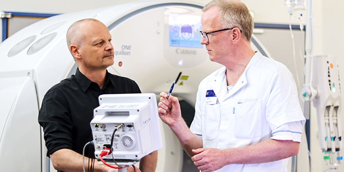
In the project, the engineers and doctors will sit together alternately at Rigshospitalet and DTU to keep a clear focus on the value of the research, explains Klaus Fuglsang Kofoed, who is a doctor in the Heart Centre at Rigshospitalet:
"The healthcare system is very concerned about whether AI is a real and pragmatic, strong partner, or - if I may say so - just a new gimmick that does not really move anything. A lot of AI gets lost in intermediate calculations, but they must never overshadow measurement parameters such as morbidity and mortality, because will it be better if we use AI? We need to take a critical approach to how we use AI so that the resources invested actually end up improving patient care. I myself am very enthusiastic about AI and hope that the project shows that it is worthwhile and that this is the way we should go.”
The math in the heart
In the project, Rigshospitalet and DTU use 12,000 cardiovascular CT scans from the unique, Danish population study The Copenhagen General Population Study, where the patients' medical history over a 10-year period is known. You know who got the two diseases - cardiac death and stroke - and therefore you can go back and see what the patient's heart looked like before one of the two diseases occurred.
"The project has the potential to help a large group of patients by being able to predict and thereby prevent cardiovascular disease, and it will be an important contribution to DTU's vision of technology for people."
Rasmus R. Paulsen, Associate Professor, Visual Computing at DTU Compute
The researchers will first develop AI-based algorithms that enable artificial intelligence to recognize the various sub-elements of the heart on CT imaging.
The risk of cardiac death is hoped to be found by examining both the shape and appearance of the muscle tissue in the heart (the myocardium), the latter by looking at the grayscale variation of the CT image inside the muscle itself. Since doctors are not entirely sure what a sick and healthy muscle looks like on a CT image, the algorithms must make a systematic description of what the muscle looks like and link it to the patient's health situation so that it can eventually be used to make a patient-specific risk assessment.
The risk of stroke is closely related to cardiac death because blood clots in the brain start in the heart, probably in the auricle, which is a kind of appendix in the left atrium. If patients get heart fibrillation (atrial fibrillation), the heart shakes, and then the blood clots can shake loose and move up into the brain. The auricle can e.g. look like a cauliflower or a chicken wing. Doctors believe that auricles resembling cauliflower have a greater risk of forming blood clots in the complicated mass that the blood can sit still inside than in auricles resembling smooth chicken wings. That thesis must also be examined by the AI model.
Proof of concept
If the project is successful, doctors will be able to use the new knowledge and use CT scans to make cardiac diagnostics, because unlike before, modern CT scans provide only a little radiation, so you can use it as a clinical screening tool without putting people at risk for cancer.
“Artificial intelligence is still very far from the clinical everyday methods when we treat patients. But AI is - as a kind of computer-assisted systematic pattern recognition - much better than the human eye at making image analyzes and can automatically measure smaller structures in the heart. If we fulfill our visions, then in the near future we can CT-scan a patient and use AI to give a much more accurate diagnosis regarding possible health risks and thus also provide more meaningful guidance to the patient to undergo a lifestyle change, receive treatment, or something else,” says Klaus Fuglsang Kofoed.
At DTU Compute, the researchers hope the research will result in a proof of concept - a good yardstick for measuring risk - to be published in scientific articles in major journals. Later, it may be implemented in a national guideline for assessing the risk of blood clots in the brain, so that it is included together with other patient data e.g. blood pressure in an overall assessment of whether the patient should be offered preventive treatment.
“In recent years, artificial intelligence has become much better at analyzing images. With advanced algorithms, today we can extract much more information from images than before to predict serious diseases. The algorithms require high quality data where patient history is known. That is why the data set is unique because as a researcher you rarely get access to so many relevant patient data,” says Rasmus R. Paulsen.
The project builds on previous collaborations, and the grant from the Novo Nordisk Foundation enables the partners to intensify development in the area.
“We are very grateful that the Novo Nordisk Foundation supports the expansion of the collaboration between Rigshospitalet and DTU. An important part of the process is to unite two very different environments. My experience is that a research environment is a fragile plant so it is important to make it run well. And once it runs, then there are unimaginable possibilities,” says Klaus Fuglsang Kofoed.
- The data set from The Copenhagen General Population Study is a population study, where data since 2003 has been collected from patients who have been examined for various diseases.
- The data set contains over 12,000 heart CT scans, where the patients' medical history over a 10-year period is known - also patients suffering from the two diseases - blood clots in the brain (stroke) and cardiac death. That makes it possible to see what the patient's heart looked like before one of the two diseases occurred.
- Artificial intelligence can analyze the CT scans and map the pattern of when a patient is at particular risk of being affected by the two diseases.
- Rigshospitalet has already collected all necessary data from scanned persons, and through the project, data will be updated further with clinical follow-up for the patients.
Learn more here