Electron microscope images provide understanding and evidence of a world we cannot see with the naked eye.
The eight electron microscopes at DTU Nanolab—Centre for Electron Nanoscopy—operate constantly from early morning until late afternoon at Lyngby Campus. Yet there is still a queue to get access to them.
Anders Baun, professor at DTU Sustain, is one of the researchers using electron microscopy images for his work. He is investigating whether nanoparticles have unwanted effects on the environment.
“It’s important for us to see how the nanoparticles organize themselves, and what happens to them when they are absorbed by a living organism. ‘Seeing is believing’. We don’t use the most sophisticated instruments at Cen. We simply need to document whether our samples contain nanoparticles, where they are, and whether they have clumped together,” says Anders Baun.
DTU is planning to expand the instrument pool, to meet the growing demand for electron microscopy—not only from DTU researchers but from research environments around the world, and to upgrade with new equipment, as the current instruments have been in service ten years.
The current focus for electron microscope development is less on achieving higher image resolution and more on increasing instrument sensitivity,” says Jakob Birkedal Wagner, Scientific Director at DTU Cen.

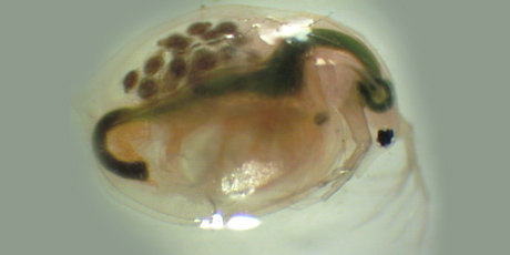
Microscopic image of an adult flea—one of the aquatic organisms used in the risk assessment of chemicals and nanoparticles. The researchers examine whether the particles simply pass through the gastrointestinal system—or are absorbed by the organism. Photo: Signe Qualmann.
“Higher sensitivity means that cameras and other detectors in the microscope get better at picking up the signal from the electron bombardment. This means that we don’t have to ‘shoot’ as many high-energy electrons at the samples we are examining, or for as long a period at a time. In other words, we are reducing the damage or changes to our samples during the actual microscope imaging process. This is particularly interesting when we are examining biological materials, which are sensitive to electron bombardment,” says Jakob Birkedal Wagner.
New electron microscopes will also make it possible to study organisms that have been fixed by freezing (cryogenic cooling). This is a field which interests Anders Baun:
“You cannot throw biological organisms into the microscope as they are. They must first be prepared, and during that process there is a risk of damaging the samples. It would therefore be interesting for us if the instruments had cryo-electron capability so that we could view the samples in their frozen state. It won’t save time, but it might provide better results.”
In addition to adding newer microscopes, DTU Cen is working on adding new laboratory facilities, to make it easier to handle biological samples in particular. The aim is for the new facilities and instruments to be ready for use in 2020.