Cancer patients can look forward to much more effective radiation treatment involving a new technology: a nano-scale marker that establishes the position of tumours in the body with pinpoint accuracy.
“Good cancer treatment demands precision, precision and precision.”
So says Professor Lena Specht, an oncology specialist at Copenhagen University Hospital (Rigshospitalet) who works with advanced radiation therapy. Lena Specht explains that when radiation is to be used to treat a cancer patient, the key consideration is to hit the tumour as accurately as possible—which can be very difficult. The reason for this is that even though an x-ray image may identify the location of the tumour, and thus the area to be bombarded with radiation, the actual position of the tumour may change during a period of treatment that can last for six weeks, for example.
“The tumour may shrink, or the surrounding tissue may become inflamed due to the treatment, making it difficult to be sure that the tumour is located exactly in the middle of the area being irradiated,” she says.
The challenge is even greater when treating patients suffering from lung cancer, because the tumour moves in time with the patient’s breathing. When, prior to treating the patient, the doctors are to define the area for irradiation, they are obliged to leave a wide margin to be sure of hitting the patient in all phases of the breathing process. In order to reduce the movement of the tumour, patients can also be asked to take a deep breath and hold it. But even then there is no way to be sure that the patient will take the same deep breath every time while the radiation therapy is underway. The doctors therefore have to irradiate a larger area to be sure of hitting the tumour, and this generates more side effects in the patient.
Today, doctors use a variety of markers to pinpoint the location of some tumours. In the case of prostate cancer, for example, they use tiny grains of gold which they place in the prostate glands of men who are to receive radiation therapy. These grains of gold are not suitable for use in lung tissue, however, because they have to be inserted using a long needle, which could easily puncture a lung and cause it to collapse.
"The idea of using a liquid that subsequently transforms into a gel is quite simply brilliant. I haven’t seen anything like it anywhere else."
Professor Lena Specht, an oncology specialist at Copenhagen University Hospital
Biomaterial injected directly into tumours
During a flight in 2010, Professor Thomas Lars Andresen from DTU Nanotech found himself sitting next to a doctor, who mentioned some of the challenges he faced regarding these markers. The DTU professor soon began to think that his knowledge of nano and biotechnology, combined with the work his team of researchers was doing in the area of biomedical engineering, might provide a solution to the problem. And the doctors in Lena Specht’s department are now conducting human trials with the solution Thomas Lars Andresen and his team came up with: a biodegradable marker that can be injected directly into the tumour.
“We’ve developed a nanostructured bio-material filled with contrast substance. This allows us to generate crystal clear images of the tumour in an x-ray scan,” explains Thomas Lars Andresen.
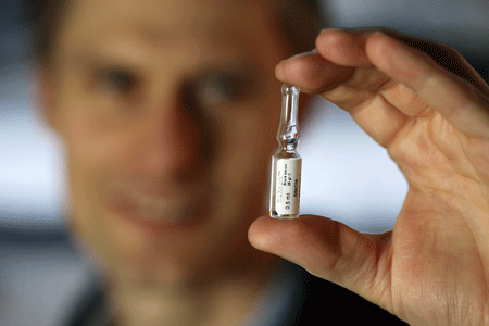
The nano-marker is in fluid form during the injection, and can therefore be inserted into the patient using an extremely thin needle. This makes it possible to work with the same equipment that is currently used to take tissue biopsies when doctors are diagnosing lung cancer. As such, doctors now have the opportunity to place the nano-marker in fragile tissue such as lung tissue. Once the fluid marker has been injected, it turns into a gel that the body itself breaks down into harmless sugars over the course of a year. Just a few microlitres are injected into the tumour, which means that the marker only extends over a couple of millimetres in the body.
|
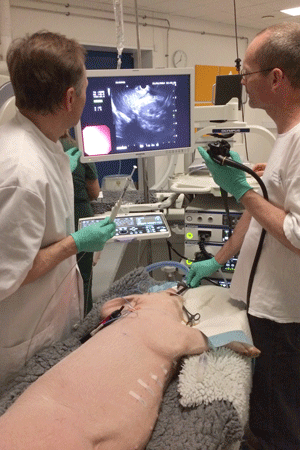
Consultant Klaus Richter Larsen (right) and Consultant Paul Clementsen (left) inserting a nano-marker in the lung of a pig.
The doctors are using ultrasound equipment to find the right position in the lung.
The marker is fluid, and it is injected using a syringe and the thin needle Paul is holding.
|
Partnership between three hospitals
It takes a great deal of experience to place the nano-marker in a lung tumour, and two consultants from Gentofte Hospital and Bispebjerg Hospital are contributing their expertise to the research project. Consultant Klaus Richter Larsen from Bispebjerg Hospital is one of them. He was involved in testing the technology in pigs and in summer 2014, he inserted the nano-marker into a human patient for the first time.
“I placed three small deposits of the nano-marker in a lung tumour. Using a bronchoscope with small ultrasound probes on the tip, I could see the tumour and the lymph nodes that had been attacked by the cancer. We use the same technology to take tissue samples when making the diagnosis,” explains Klaus Richter Larsen.
Once the nano-marker is in place, the patient is sent for radiation therapy as planned at Rigshospitalet, where Lena Specht’s department uses the marker to generate the clear images of the extent and placement of the cancerous tumour.
Over the coming year, the doctors expect to place nano-markers in 15–20 patients with lung cancer.
Brilliant solution
With clear images and neatly defined irradiation areas in the patients, doctors can control the doses of radiation much more accurately, ensuring that they have maximum impact on the tumour while avoiding healthy tissue as far as possible. “As we can irradiate more accurately, we can ramp up the dosage without the patient suffering more side effects or consequential damage to healthy tissue. This should allow us to cure more patients with tumours, and we should also be able to treat larger tumours, which we cannot currently irradiate,” says Lena Specht.
According to the professor, the technology has potential for additional areas of use in the future:
"In patients with oesophageal cancer, it can be difficult to see on a scan where the tumour starts and where it ends. With a marker at each end, however, we would be able to irradiate much more accurately along the full length of the tumour. I am also fairly sure that we could use the marker in treating breast cancer. We currently use needles to indicate where the surgeon has to go in to remove a node. We may be able to use nano-markers instead in future,” says Lena Specht.
Lena is thrilled with the new technology.
“Over the years, I have assessed many other projects involving markers. However, they all features solutions based on metal, which shows up clearly on x-rays. The only problem is that metal markers have to be inserted using thick, rigid needles that often cause damage such as perforations to the surrounding tissue. The idea of using a liquid that subsequently transforms into a gel is quite simply brilliant. I haven’t seen anything like it anywhere else.”
Article from DYNAMO no. 38, DTU's quarterly magazine.
An American journal has highlighted the DTU spin-out Nanovi as one of the most promising players in the field of radiation therapy.
Nanovi is a spin-out from DTU Nanotech. The company produces the tumour marker BioXmark, which is based on research conducted by Professor Thomas Lars Andresen and his team.
In spring 2014, Nanovi was selected and presented as one of the most promising start-up enterprises focusing on radiation therapy for cancer. The profile was published in the American journal Start-up, which specializes in in-depth analyses and reviews of new enterprises for the benefit of investors, business developers and others.
BioXmark is a biodegradable tumour marker which, filled with contrast fluid, can be injected into cancerous growths in patients to help generate crystal clear images of the tumours through x-ray scanning. The results can be used to increase the precision of radiation therapy, thus improving the treatment of cancer patients and reducing side-effects.